 English
 中文
中文 Français
 Español
 Pусский
What's ultrasonography ? ultrasound pregnancy imaging, diagnostic ultrasound imaging system
Defination of Ultrasonography
The terms “ultrasound” ,"ultrasound pregnancy imaging", “ultrasonography” and “sonogram” may sound familiar to you. Widely used in the last 50 years, doctors first applied ultrasonography in medicine to detect pregnancy. In a frame or scrapbook at home, you may even have your child’s first “sonogram.”
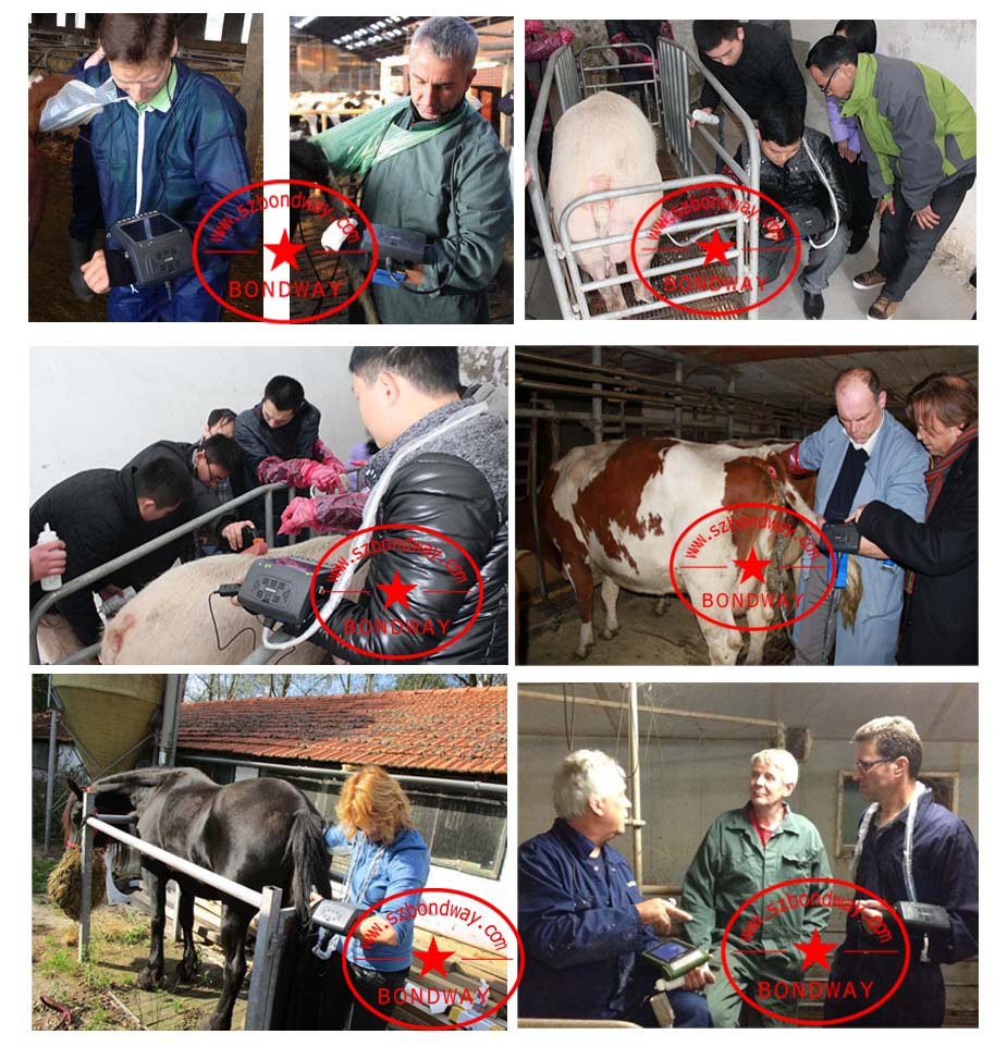
A sonogram is the image created on a screen or print out produced by a medical ultrasound. Just like human medicine, veterinarians first used ultrasound in to detect pregnancy in animals, however, they now use it for a wide variety of reasons.
The term “ultrasound” refers to sound waves with a frequency too high for humans to hear, not unlike the silent dog whistle you use to train Buster. An ultrasound wave produces an image when sent through the body with the use of a probe. The sound echoes off parts of the tissue and then displays a sonogram on the screen.
Ultrasonography is a very valuable diagnostic procedure. Radiographs show us the general shapes of structures, such as the size and shape of the heart or liver, but ultrasound allows us to look at the inside architecture of our major organs (heart, liver, gall bladder, pancreas, kidneys, urinary bladder, stomach, intestines and adrenal glands). It also allows us to evaluate the function of the heart, as we can view the movement and size of the heart walls, chambers and heart valves.
Having several advantages over other forms of diagnostic imaging, ultrasonography shows a live image, and diagnosis can be immediate. You will generally know the results of an ultrasound soon after our practice reviews the data gathered. However, ultrasound cannot view gas filled or bony tissue and other forms of diagnostic imaging like a CT scan may better view certain areas of the body.
Another common kind of ultrasonography is the Echocardiogram. Echocardiography evaluates the heart. It can view and carefully evaluate movement and size of the walls, chambers and valves of the heart.
Anesthesia is not necessary; however sedation is not uncommon. Because ultrasound waves cannot transmit through air, our practice shaves your pet’s fur to provide direct contact to the skin. A water-soluble gel helps with conduction and may feel slightly wet or cold, but the procedure shouldn’t feel uncomfortable. There are no documented risks and no ionizing radiation is involved.
Shenzhen Bondway Electronics Co. Ltd
2017-10-16
Best sellers
-
 Digital Vet Ultrasound scanner for equine, bovine, cattle, llama
-
 Veterinary ultrasound machine for equine, bovine,camel
-
 Digital Palmtop veterinary ultrasound scanner (Discontinued since April 29, 2022)
-
 Digital veterinary ultrasound for farm animals
-
 Digital Veterinary Ultrasound Scanner-ultrasound scan for swine, obvine,goat,alpacca
-
副本.png?imageView2/2/w/200/h/150/q/60) Digital color doppler ultrasound imaging system
Digital color doppler ultrasound imaging system
-
 Digital color doppler ultrasound imaging system
-
 Digital color doppler ultrasound imaging system


副本.png?imageView2/2/w/200/h/150/q/60)